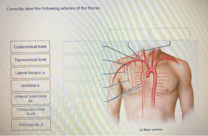
Correctly Label the Following Arteries of the Thorax
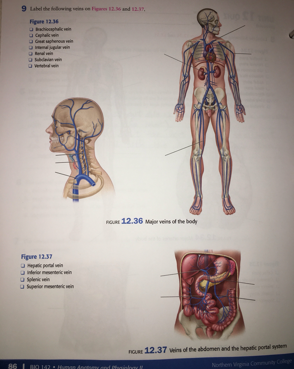
Theyre present only in the upper 9 spaces and every space includes 2 veins and accompanies the anterior intercostal arteries. All the thoracic arteries originate from the aorta and the three largest ones are the brachiocephalic trunk left common carotid artery and left subclavian artery.

Circulatory Pathways Anatomy And Physiology Ii
Page 12 of 20 StudyBlue printing of Chapter 21.

. External iliac artery- Lower limb 8. Correctly label the following arteries of the abdomen and pelvic region. Correctly label the following anatomical features of the thoracic cavity.
Respond to the questions in the pop-up boxes addressing the structure of the wall of. Right gastric artery- Stomach 3. Supreme thoracic artery supplies blood to the upper chest.
Its only branches are the coronary arteries. The ascending aorta rises for about 5 cm above the left ventricle. Suprarenal artery -Adrenal gland 5.
Correctly label the following veins of the thorax. Blood vessels are the pipes or channels through through blood flows and reaches to. Thoracoacromial trunk supplies blood to the deltoid and pectoral region.
All systemic arteries arise from the aorta which has three principal regions. Anterior Intercostal Veins. Correctly label the following structures related to the position of the heart in the thorax.
Adjust credit for all students. Lateral thoracic artery supplies blood to the lateral thorax chest wall. Score answer.
The anterior and posterior interventricular sulci overlie an internal wall the interventricular septum that divides the right ventricle from the left. Renal artery- Kidney 6. Blood Vessel Circulation 2020-02-18.
The pathway labeled _____ shows alternative routes of blood supply found in the heart. Median sacral artery -Inferior vertebrae 7. The coronary sulcus and two interventricular sulci harbor the largest of the coronary blood vessels.
Correctly label the following veins of the abdomen and pelvic region. Your Skills Rank. The heart is located in the thoracic cavity in the mediastinum between the lungs and deep to the sternum.
Worksheet labeling quiz for arteries and veins cat arteries diagram upper arteries of a cat youtube title cat 2 18. Internal carotid a Costocervical trunk Thyrocervical trunk Prev 10 of 26 B 11. Correctly label the following internal anatomy of the heart.
Correctly label the following veins of the thorax. Correctly label the following coronary blood vessels of the heart. There are 2 common carotid arteries.
Correctly label the following veins of the thorax. From the quiz author. Hepatic artery- Liver 4.
Subscapular artery supplies blood to the muscles of the scapula and latissimus dorsi. Correctly label the following features of the aorta and its major branches. Note that the human heart obtains a substantial amount of its energy from fats.
Study quiz for Anatomy and Physiology This quiz has tags. Anatomy and Physiology questions and answers. View Screenshot 20png from BIO 1400 at CUNY Kingsborough Community College.
Correctly label the following features of the aorta and its major branches. Bronchial artery- Lung tissue 2. Dissection veins and arteries quiz bing can you name the arteries of cat you re not logged in compare scores with friends on all sporcle quizzes name the vein which Label the major arteries and veins.
The anterior side typically contains more adipocytes than the posterior side. If you take a close look at the previous table and diagram you will see that several major arteries and veins travel through the thorax. Correctly label the following parts of the pericardium and the heart walls.
The aortic arch curves to the left like an inverted U superior to. Label the components of the cardiac conduction system. Correctly label the following arteries of the thorax.
This is an online quiz called Arteries of the Thorax. Major Arteries of the Body. Click on the tags below to find other quizzes on the same subject.
Identify the following structures of the heart. Correctly label the following anatomical features of the thoracic cavity. They conclude in the upper 6 spaces they stop in the internal thoracic vein and in seventh eighth and ninth spaces they stop in the musculophrenic vein.
This figure shows variations in circulatory pathways. Correctly label the following pulse points. Inferior mesenteric artery- Descending colon and rectum.
-Book Print Carotid sinus External carotid a Common carotid a. Several visceral arteries also supply various thoracic organs including. The right common carotid artery originates behind the sternoclavicular joint in neck from brachiocephalic trunk innominate arteryThe left common carotid artery originates directly from the arch of aorta in thorax superior mediastinum.
Internal thoracic artery Arteria thoracica interna The internal thoracic artery internal mammary artery is a long paired vessel that originates from the proximal part of the subclavian arteryIt runs inferomedially and enters the thoracic cage deep to the clavicle and the first ribTerminating at the level of the sixth rib it divides into two terminal branches. Blood vessels are the pipes or channels through through blood flows and reaches to. From its superior to inferior midpoints it is tilted toward the left so about twothirds of the heart lies to the left of the median plane.
Drag each label into the appropriate position to identify the waves of a normal ECG. Correctly label the following arteries of the head and neck. Correctly label the following arteries of the thorax.
Solved Correctly Label The Following Arteries Of The Thorax Chegg Com
19 5 Heart Position Within The Thoracic Cavity A The Heart Is Located Within The Mediastinum Of The Thoracic Cavity Betwee Thoracic Cavity Thoracic Cavities
Answered Label The Following Arteries In Figures Bartleby
No comments for "Correctly Label the Following Arteries of the Thorax"
Post a Comment